Products

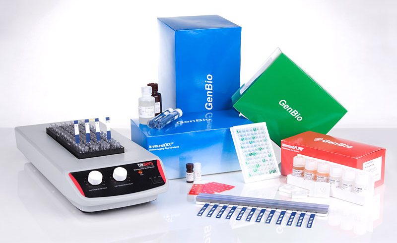
ImmunoDOT
The ImmunoDOT Test utilizes an enzyme-linked immunoassay (EIA) dot technique for the detection of antibodies. The antigens are dispensed as discrete dots onto a solid membrane. After adding specimen to a reaction vessel, an assay strip is inserted, allowing patient antibodies reactive with the test antigen to bind to the strip’s solid support membrane. In the second stage, the reaction is enhanced by removal of non-specifically bound materials. During the third stage, alkaline phosphatase-conjugated anti-human antibodies are allowed to react with bound patient antibodies. Finally, the strip is transferred to enzyme substrate reagent, which reacts with bound alkaline phosphatase to produce an easily seen, distinct dot.
ImmunoFA
The ImmunoFA Test utilizes the indirect fluorescent antibody technique for the detection and titration of antibody in human sera. The antigen substrate is dried on microscope slides. The organisms are fixed and no infective forms can be detected using in vivo inoculation methods. Test serum is applied to the antigen substrate and incubated at 37 °C. Following incubation, the serum is rinsed from the slide and fluorescein-conjugated antihuman antibody is applied. Following a second incubation, the slide is rinsed and examined under a fluorescence microscope. If antibody to the organism is present in the test serum, it will combine with the antigens of the fixed organisms and the fluorescein-conjugated anti-antibody will be bound causing the organisms to fluoresce. The reaction is considered positive when the majority of the fixed organisms exhibit fluorescence around their entire periphery.
ImmunoFLOW
ImmunoFLOW is an immunoassay consisting of a cassette and three reagents. The cassette contains a paper matrix (for example, nitrocellulose) and an absorbent material. The paper matrix was manufactured with three “dots”, each contain an antigen (e.g., positive control, analyte 1, analyte 2). A body fluid (e.g., serum) is applied to the triangular opening and allowed to flow through the paper matrix into the absorbent. To assure assay specificity, a wash reagent is applied and flows into the absorbent material. Finally, gold particles attached to an immunological reagent (e.g., anti-immunoglobulin) is applied and absorbed. The “dot” applied to the paper matrix contains antigen. Specific antibody in body fluid will bind to the antigen. If specific antibody binding occurs, immunologically active gold particles will bind and cause a red/pink color formation.
ImmunoWELL
The ImmunoWELL Test utilizes an EIA microtiter plate technique for the detection of antibodies. Antihuman IgG treated serum is added to antigen coated microtiter wells and allowed to react. After removal of unbound antibodies, horseradish peroxidaseconjugated antihuman IgM antibodies are allowed to react with bound antibodies. The bound peroxidase reacts with 2,2′-azinodi-[3-ethylbenzthiazoline sulfonate] (ABTS®), the chromogenic substrate, developing a color. Finally, the substrate reaction is stopped and the optical density is read with a spectrophotometric microwell reader.
